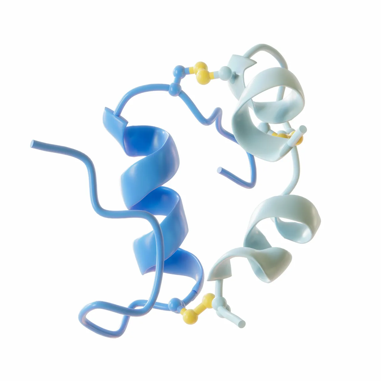
Insulin struct
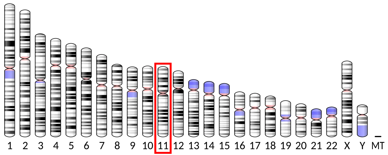
Chromosome 11 (human)
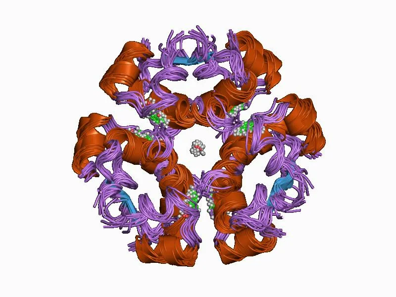
1aiy: R6 HUMAN INSULIN HEXAMER (SYMMETRIC), NMR, 10 STRUCTURES
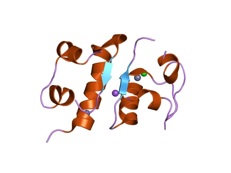
1fub: FIRST PROTEIN STRUCTURE DETERMINED FROM X-RAY POWDER DIFFRACTION DATA

1qiy: HUMAN INSULIN HEXAMERS WITH CHAIN B HIS MUTATED TO TYR COMPLEXED WITH PHENOL

2bn1: INSULIN AFTER A HIGH DOSE X-RAY BURN
Insulin
Insulin is a peptide hormone produced by beta cells of the pancreatic islets encoded in humans by the insulin (INS) gene. It is the main anabolic hormone of the body.
Venomous Snails Weaponizing Metabolic Shock
Predatory cone snails like Conus geographus and Conus tulipa do not rely exclusively on traditional neurotoxins to immobilize prey. Instead, they synthesize modified versions of insulin within their venom cocktails. This weaponized hormone mimics the molecular structure of fish insulin rather than the snail's own variety. When injected into the water or prey, it causes rapid hypoglycemic shock, slowing down fish for capture.
From Wikipedia · CC BY-SA 4.0

The chemical structure of testosterone.
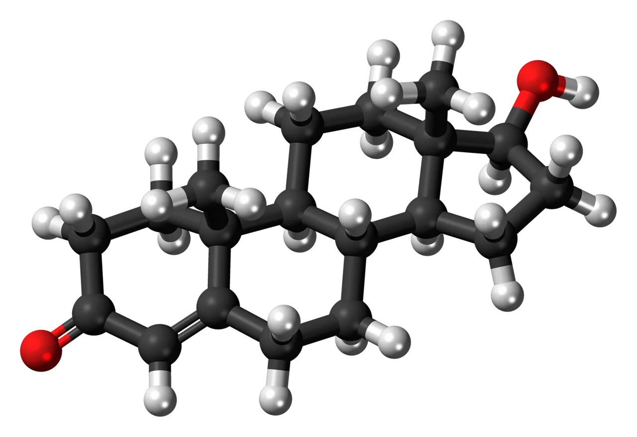
A ball-and-stick model of testosterone.
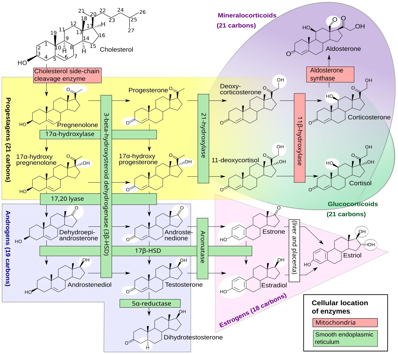
.mw-parser-output .vanchor>:target~.vanchor-text{background-color:#ebf4ff}@media screen{html.skin-theme-clientpref-night .mw-parser-output .vanchor>:target~.vanchor-text{background-color:#0f4dc9}}@media screen and (prefers-color-scheme:dark){html.skin-theme-clientpref-os .mw-parser-output .vanchor>:target~.vanchor-text{background-color:#0f4dc9}}Figure 1: Human steroidogenesis, showing testosterone near bottom[29]
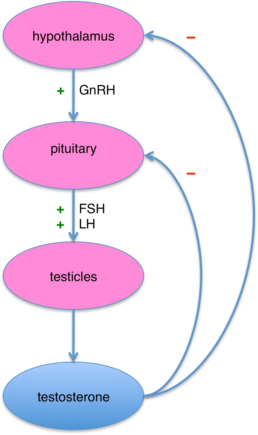
Figure 2. Hypothalamic–pituitary–testicular axis
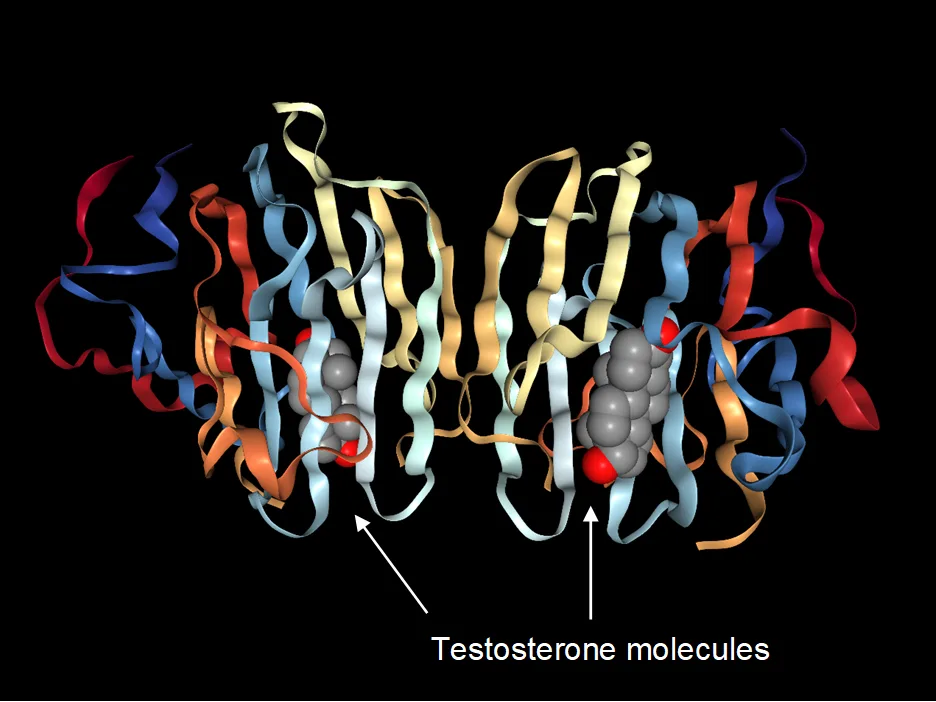
Dimeric sex hormone-binding globulin with its testosterone ligands
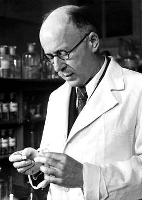
Nobel Prize winner, Leopold Ružička of Ciba, a pharmaceutical industry giant that synthesized testosterone
Testosterone
Testosterone is the primary male sex hormone and androgen in males, driving the development of male reproductive tissues and secondary sexual characteristics like increased muscle and bone mass.
Estrogen as the Architect of Male Brains
Although estrogen is widely considered the quintessential female hormone, it directly directs male neural development during early development. Circulating testosterone crosses the blood–brain barrier into the male fetal brain, where aromatization transforms it into estradiol to drive masculinization. Female fetuses avoid this neural fate because α-fetoprotein binds circulating estrogen, preventing the hormone from altering female brain structure.
From Wikipedia · CC BY-SA 4.0

Estradiol
![Reference ranges for the blood content of estradiol, the primary type of estrogen, during the menstrual cycle[18]](https://nwjvzop1xgplmzvt.public.blob.vercel-storage.com/wikifeed/estrogen/1-ea6831a386.webp)
Reference ranges for the blood content of estradiol, the primary type of estrogen, during the menstrual cycle[18]
![Steroidogenesis, showing estrogens at bottom right as in pink triangle[112]](https://nwjvzop1xgplmzvt.public.blob.vercel-storage.com/wikifeed/estrogen/2-9a2aaa56ee.webp)
Steroidogenesis, showing estrogens at bottom right as in pink triangle[112]
Estrogen
Estrogen is a category of sex hormone responsible for the development and regulation of the female reproductive system and secondary sex characteristics.
The Hidden Prerequisite for Male Libido
While commonly viewed as an exclusively female hormone, estrogen plays an essential role in male physiology and sexual motivation. Libido depends on androgen levels only when estrogens are present to facilitate desire. Without estrogen, circulating free testosterone actually decreases sexual desire rather than increasing it. In males, estrogen also regulates crucial reproductive functions necessary for the maturation of sperm.
From Wikipedia · CC BY-SA 4.0

The chemical structure of progesterone
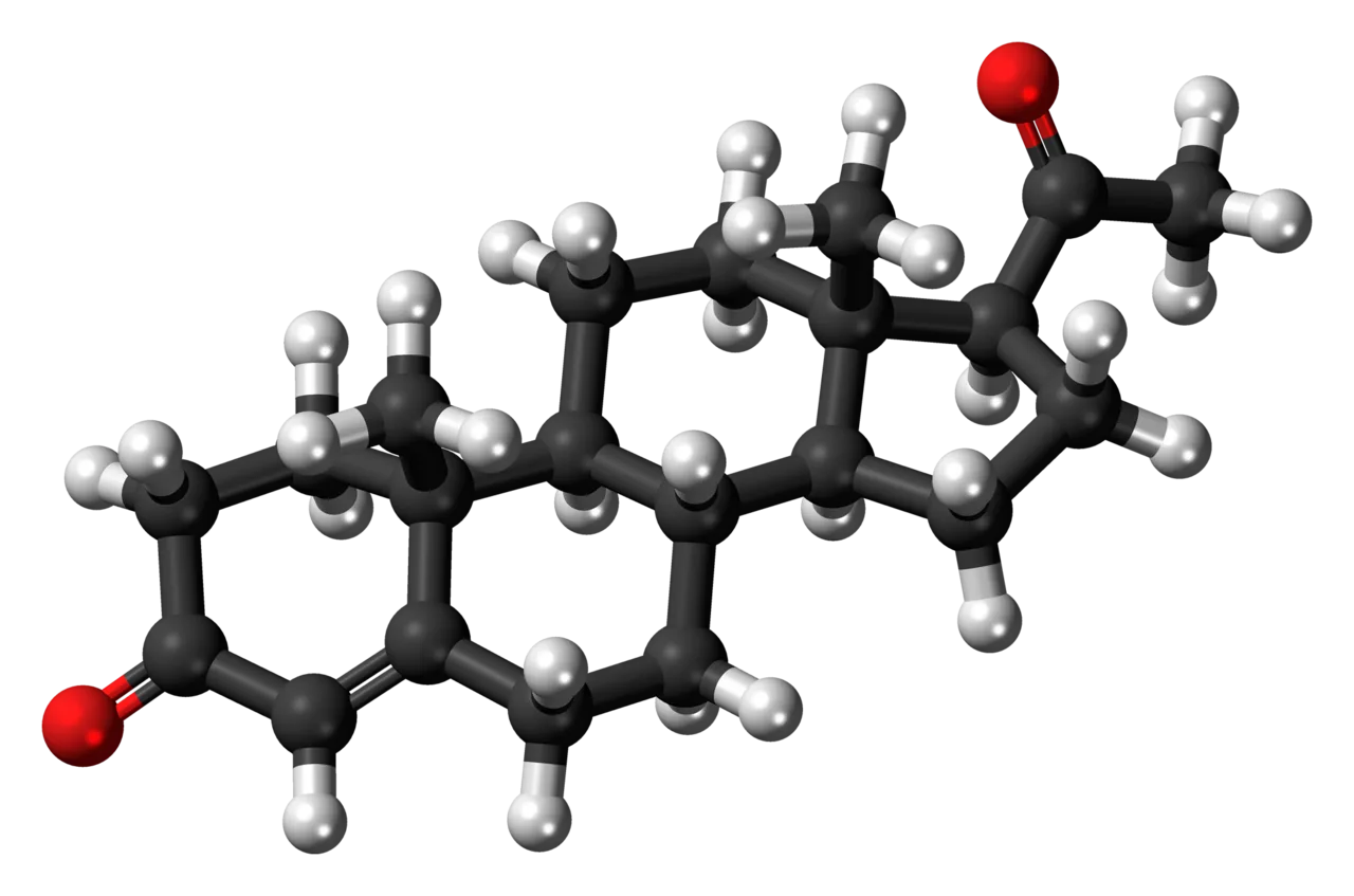
A ball-and-stick model of progesterone.
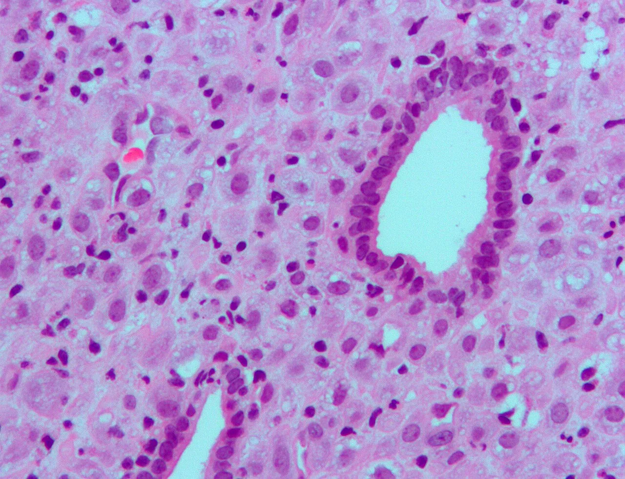
Micrograph showing changes to the endometrium due to progesterone (decidualization) H&E stain
![Progesterone levels across the menstrual cycle in normally cycling and ovulatory women.[132] The horizontal lines are th](https://nwjvzop1xgplmzvt.public.blob.vercel-storage.com/wikifeed/progesterone/3-d4ca64e37c.webp)
Progesterone levels across the menstrual cycle in normally cycling and ovulatory women.[132] The horizontal lines are the mean integrated levels for each curve; the vertical line is mid-cycle.
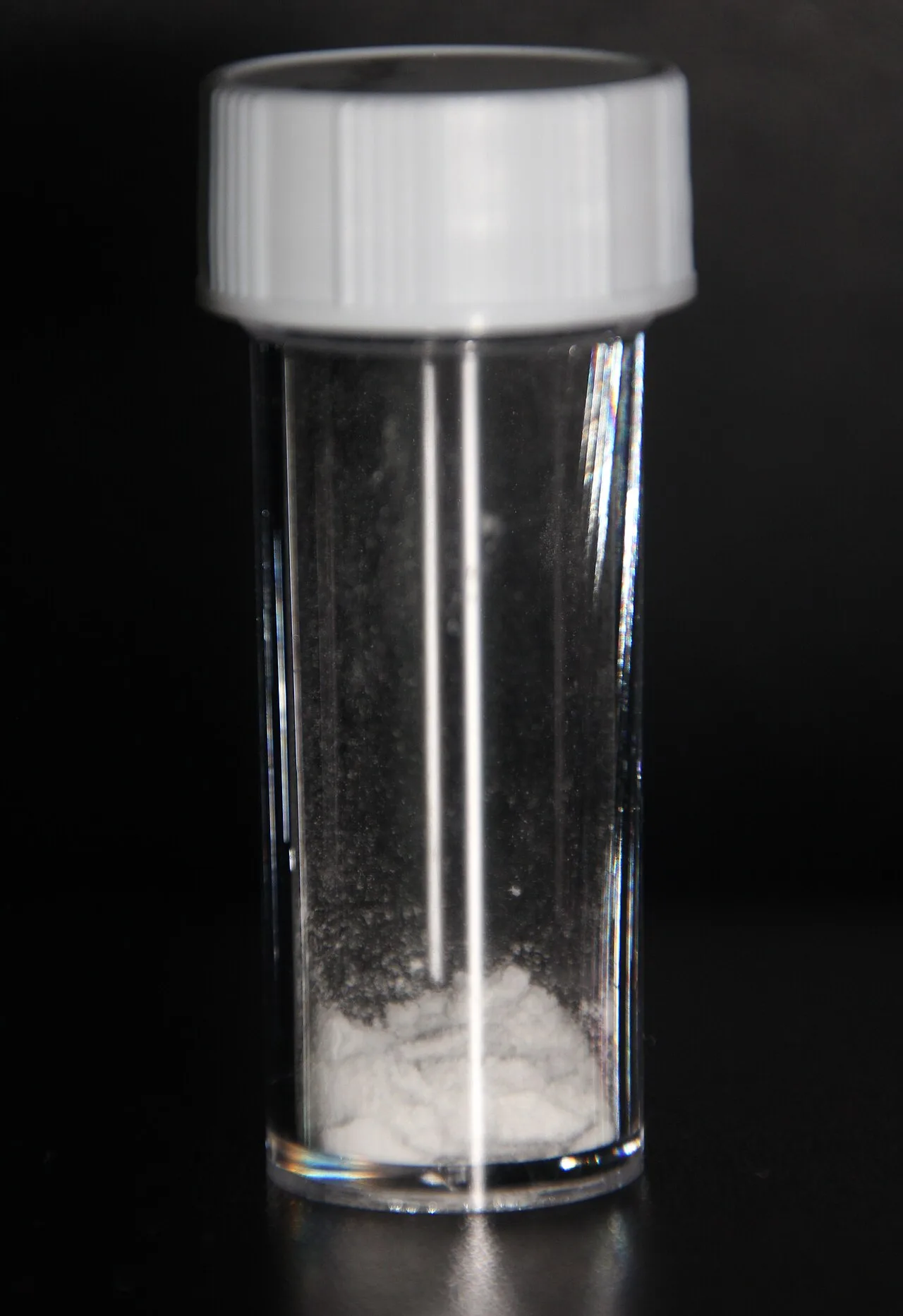
A sample of progesterone
![Stigmasterol to progesterone synthesis[166][167][168][169][170]](https://nwjvzop1xgplmzvt.public.blob.vercel-storage.com/wikifeed/progesterone/5-bbe114813a.webp)
Stigmasterol to progesterone synthesis[166][167][168][169][170]
Progesterone
Progesterone (P4) is an endogenous steroid and progestogen sex hormone involved in the menstrual cycle, pregnancy, and embryogenesis of humans and other species.
Fueling Male Genital Differentiation
Commonly viewed strictly as a female reproductive hormone, progesterone is an indispensable precursor for male fetal development. Placental progesterone converts into 5α-dihydrotestosterone (DHT), the potent androgen required for the formation of male genitalia. This transformation occurs both through direct intermediate conversion to testosterone and via the specialized androgen backdoor pathway.
From Wikipedia · CC BY-SA 4.0
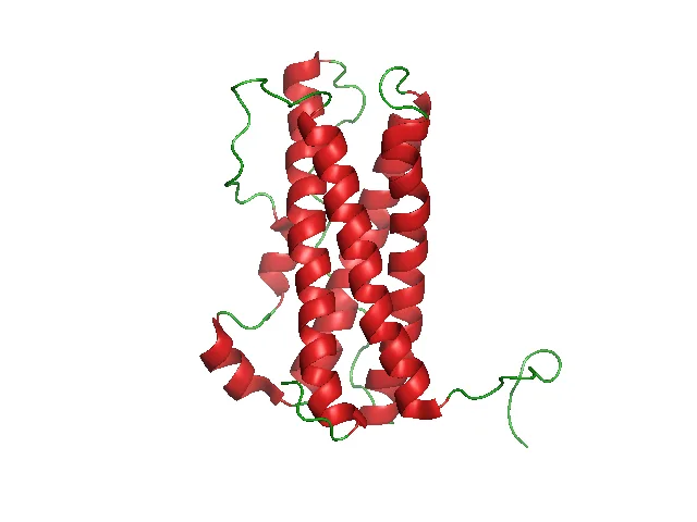
PRL structure
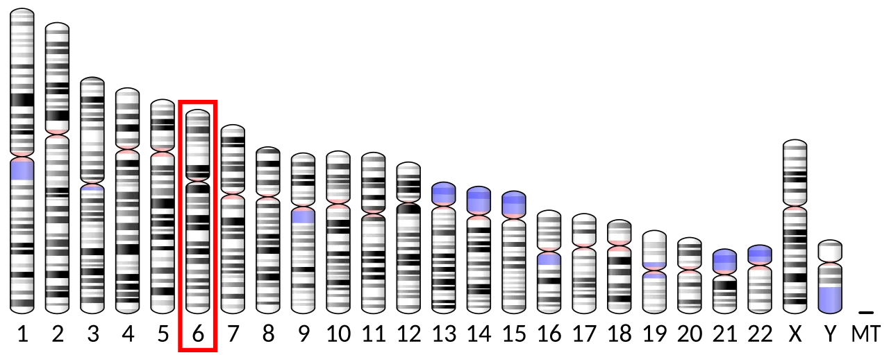
Chromosome 6 (human)

Chromosome 6 (human)
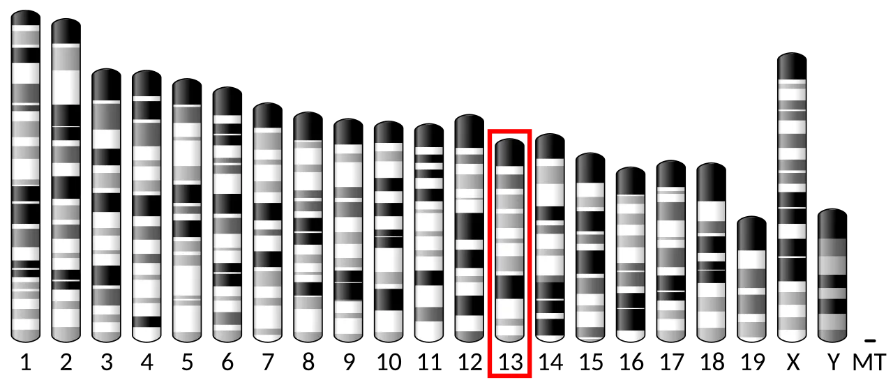
Chromosome 13 (mouse)

Chromosome 13 (mouse)
Prolactin
Prolactin is a protein best known for its role in enabling mammals to produce milk. Secreted from the pituitary gland, it is influential in over 300 processes in various vertebrates.
Default Inhibition in the Pituitary
Most pituitary hormones require an active biochemical trigger to stimulate their release into systemic circulation. Prolactin operates under an inverted regulatory logic: it is the only anterior pituitary hormone whose principal control is inhibitory. Neurosecretory tuberoinfundibulum (TIDA) neurons in the arcuate nucleus continually release dopamine to engage lactotroph D2 receptors. When this dopaminergic brake is interrupted, pituitary prolactin secretion is permitted to surge.
From Wikipedia · CC BY-SA 4.0
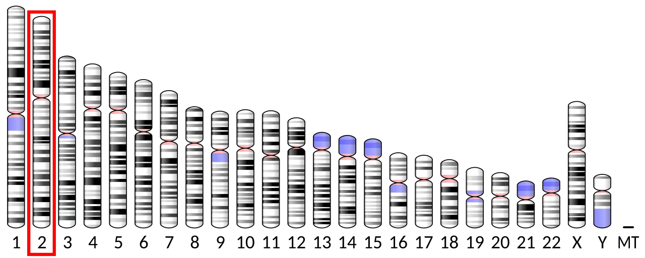
Chromosome 2 (human)

Chromosome 2 (human)
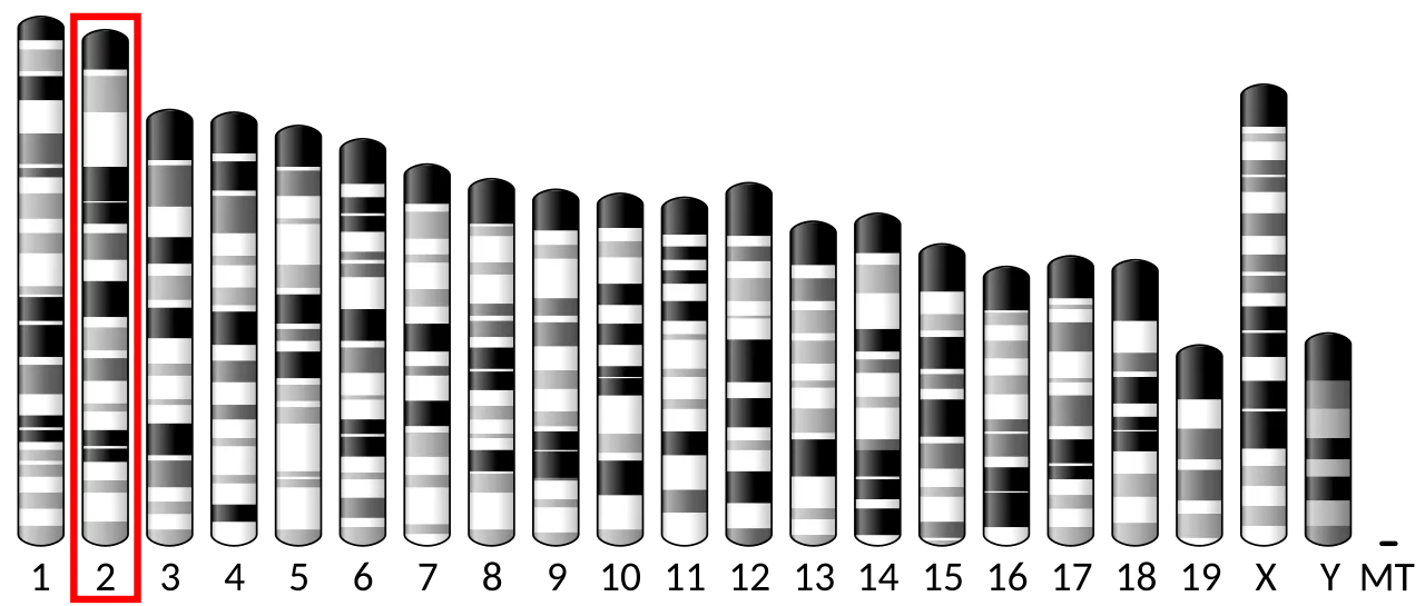
Chromosome 2 (mouse)

Chromosome 2 (mouse)
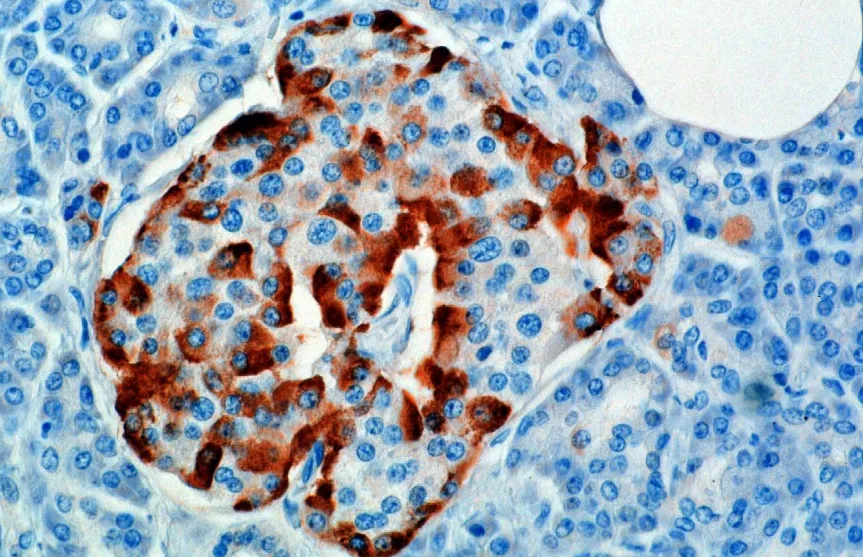
A microscopic image stained for glucagon
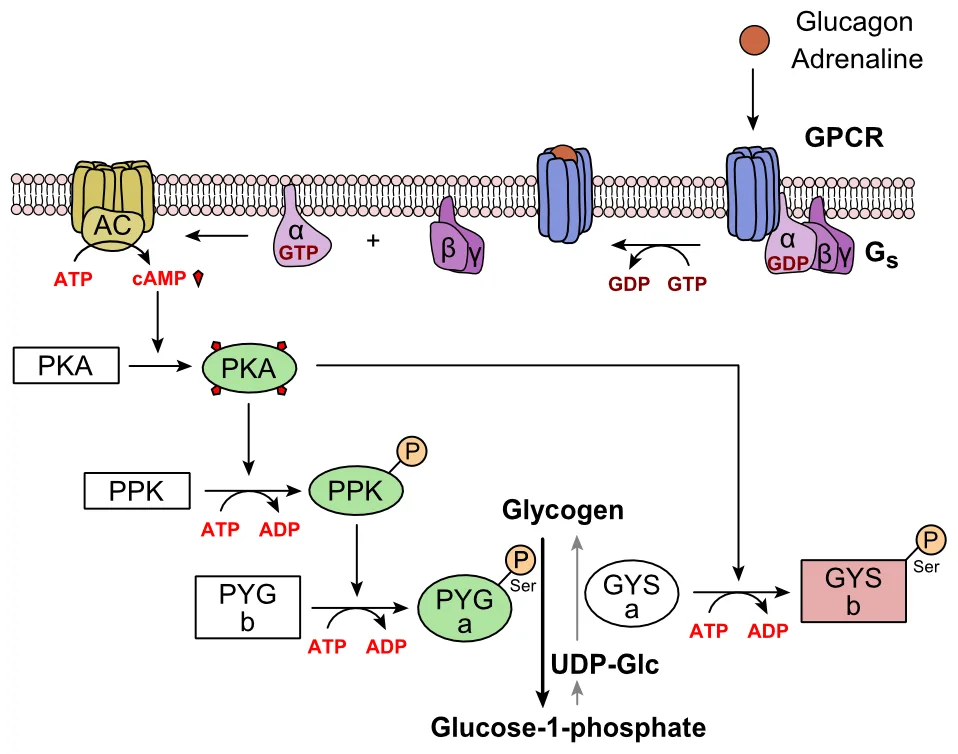
Metabolic regulation of glycogen by glucagon.
Glucagon
Glucagon is a peptide hormone produced by alpha cells of the pancreas that raises the concentration of glucose and fatty acids in the bloodstream.
Anglerfish Islets and the Road to GLP-1
Major breakthroughs in modern diabetes medicine trace back to marine biology. In 1979, researchers harvested Brockman bodies from American anglerfish to study hormone genetics in bacterial cultures. By 1982, P. Kay Lund and Richard Goodman demonstrated that anglerfish proglucagon encoded three distinct peptides. This milestone enabled Graeme Bell's team to isolate glucagon-like peptide-1, creating the foundation for GLP-1 receptor agonists.
From Wikipedia · CC BY-SA 4.0

Thyroxine2
![Biosynthesis of thyroxine[4]](https://upload.wikimedia.org/wikipedia/commons/thumb/0/0f/Thyroxine_biosynthesis.svg/1280px-Thyroxine_biosynthesis.svg.png?utm_source=en.wikipedia.org&utm_campaign=imageinfo&utm_content=thumbnail)
Biosynthesis of thyroxine[4]
Thyroxine
Thyroxine, also known as tetraiodothyronine or T4, is a hormone produced by the thyroid gland that serves as the primary form of thyroid hormone in the blood and regulates metabolic rate.
A Circulating Prohormone Reservoir
Although thyroxine is the predominant thyroid hormone circulating throughout the bloodstream, it primarily acts as a prohormone. It serves as the direct precursor to triiodothyronine (T3), which wields greater biological activity. Together with its active metabolites, thyroxine maintains metabolic rate, promotes brain development, and preserves heart, muscle, and bone health.
From Wikipedia · CC BY-SA 4.0
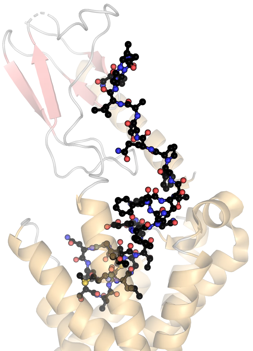
Human calcitonin bound to receptor PDB 7TYO
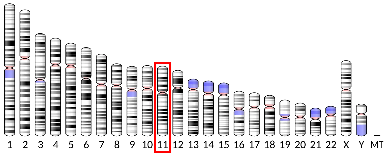
Chromosome 11 (human)

Chromosome 11 (human)

Chromosome 7 (mouse)

Chromosome 7 (mouse)

Structure of human calcitonin receptor-Gs complex. The transmembrane calcitonin receptor (blue) is bound to human calcitonin (red) and the Gs complex (yellow). PDB: 7TYO
Calcitonin
Calcitonin is a 32-amino-acid peptide hormone secreted by thyroid parafollicular cells that acts to reduce blood calcium, opposing the effects of parathyroid hormone.
A Misidentified Glandular Origin
Douglas Harold Copp and B. Cheney first purified calcitonin in 1962 at the University of British Columbia. Medical researchers initially assumed the hormone was secreted by the parathyroid gland. However, Iain Macintyre and his team at the Royal Postgraduate Medical School in London proved that thyroid parafollicular cells are the true origin. This corrected early assumptions and grounded modern calcium endocrinology.
From Wikipedia · CC BY-SA 4.0

Protein CGA PDB 1dz7

Effects of LH on the body
![Reference ranges for the blood content of luteinizing hormone (LH) during the menstrual cycle.[31] The ranges denoted By](https://upload.wikimedia.org/wikipedia/commons/thumb/c/ca/Luteinizing_hormone_%28LH%29_during_menstrual_cycle.png/1280px-Luteinizing_hormone_%28LH%29_during_menstrual_cycle.png?utm_source=en.wikipedia.org&utm_campaign=imageinfo&utm_content=thumbnail)
Reference ranges for the blood content of luteinizing hormone (LH) during the menstrual cycle.[31] The ranges denoted By biological stage may be used in closely monitored menstrual cycles in regard to other markers of its biological progression, with the time scale being compressed or stretched to how much faster or slower, respectively, the cycle progresses compared to an average cycle.The ranges denoted Inter-cycle variability are more appropriate to use in non-monitored cycles with only the beginning of menstruation known, but where the woman accurately knows her average cycle lengths and time of ovulation, and that they are somewhat averagely regular, with the time scale being compressed or stretched to how much a woman's average cycle length is shorter or longer, respectively, than the average of the population.The ranges denoted Inter-woman variability are more appropriate to use when the average cycle lengths and time of ovulation are unknown, but only the beginning of menstruation is given.

Lateral flow test strip for urine LH, used to predict ovulation
![Chance of pregnancy by menstrual cycle day of insemination relative to ovulation[35]](https://nwjvzop1xgplmzvt.public.blob.vercel-storage.com/wikifeed/luteinizing-hormone/4-ebd19c605b.webp)
Chance of pregnancy by menstrual cycle day of insemination relative to ovulation[35]
Luteinizing hormone
Luteinizing hormone is a hormone produced by gonadotropic cells in the anterior pituitary gland. In females, an acute rise of LH triggers ovulation and the development of the corpus luteum.
Latin Yellows and the Ovarian Transformation
The designation of luteinizing hormone traces directly to the Latin word luteus, meaning yellow. This moniker reflects its role in developing the corpus luteum, a cellular ovarian mass with a distinct yellow pigmentation that forms after egg release. Rather than describing the hormone molecule itself, the name immortalizes the colorful physical structure left behind in the ovary after luteinization occurs.
From Wikipedia · CC BY-SA 4.0
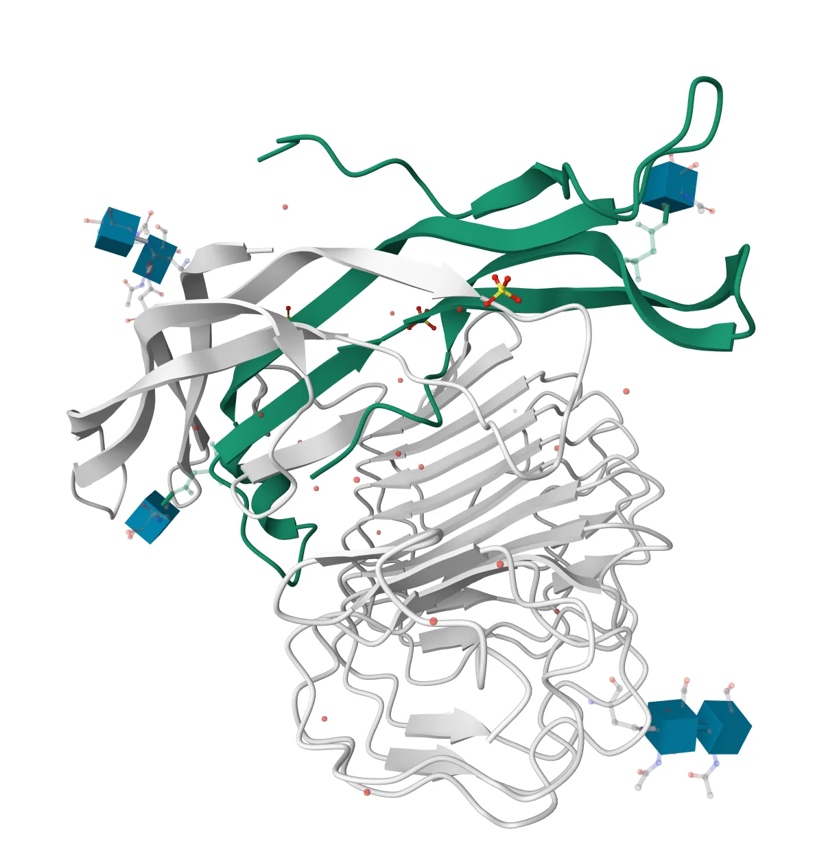
Glycoprotein hormones alpha chain within Follicle Stimulating Hormone and Receptor complex 1XWD
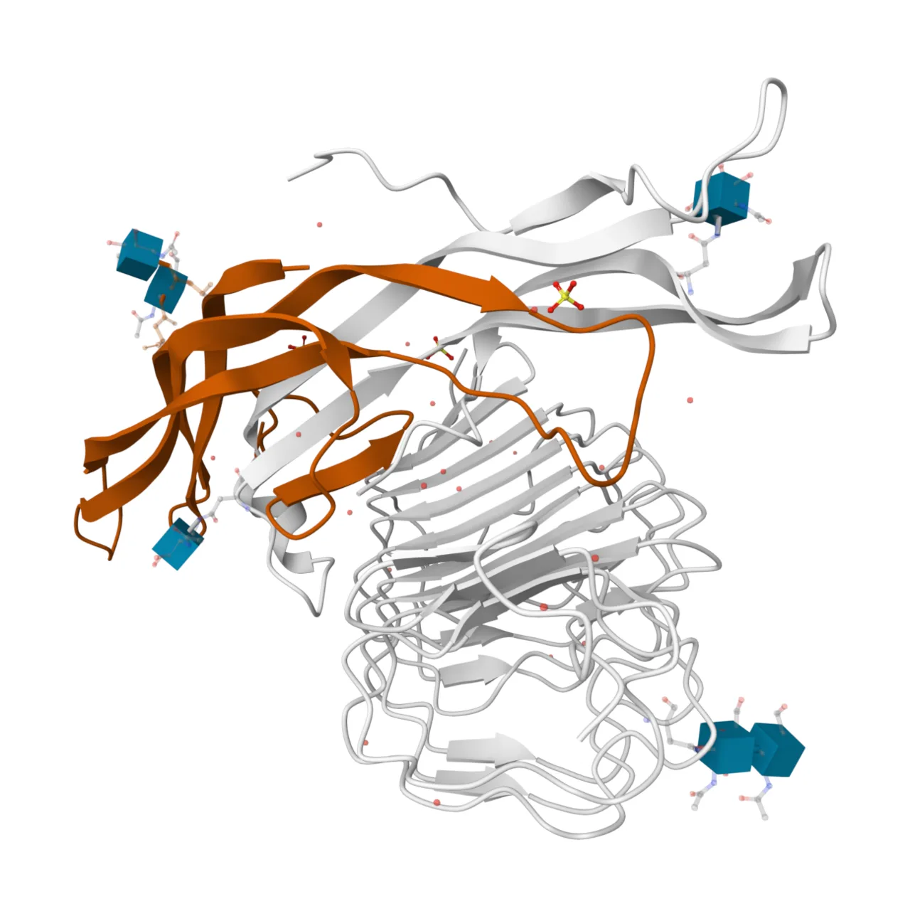
Follitropin subunit beta within Follicle Stimulating Hormone and Receptor complex 1XWD
![Reference ranges for the blood content of follicle-stimulating hormone levels during the menstrual cycle.[12] The ranges](https://nwjvzop1xgplmzvt.public.blob.vercel-storage.com/wikifeed/follicle-stimulating-hormone/2-fff1e2d8cd.webp)
Reference ranges for the blood content of follicle-stimulating hormone levels during the menstrual cycle.[12] The ranges denoted By biological stage may be used in closely monitored menstrual cycles in regard to other markers of its biological progression, with the time scale being compressed or stretched to how much faster or slower, respectively, the cycle progresses compared to an average cycle.The ranges denoted Inter-cycle variability are more appropriate to use in non-monitored cycles with only the beginning of menstruation known, but where the woman accurately knows her average cycle lengths and time of ovulation, and that they are somewhat averagely regular, with the time scale being compressed or stretched to how much a woman's average cycle length is shorter or longer, respectively, than the average of the population.The ranges denoted Inter-woman variability are more appropriate to use when the average cycle lengths and time of ovulation are unknown, but only the beginning of menstruation is given.
Follicle-stimulating hormone
Follicle-stimulating hormone (FSH) is a glycoprotein polypeptide hormone that regulates the development, growth, pubertal maturation, and reproductive processes of the body.
A Shared Peptide Backbone Across Distinct Hormones
Four distinct hormones—FSH, LH, TSH, and hCG—share an identical alpha subunit composed of 96 amino acids. Biological specificity is conferred entirely by the variable beta subunit, which spans 111 amino acids in FSH. This modular structure demonstrates how endocrine systems repurpose a universal protein scaffold while delegating receptor targeting to a specialized companion chain.
From Wikipedia · CC BY-SA 4.0
![The system of the thyroid hormones T3 and T4.[4]](https://nwjvzop1xgplmzvt.public.blob.vercel-storage.com/wikifeed/thyroid-stimulating-hormone/0-8d046b2e6b.webp)
The system of the thyroid hormones T3 and T4.[4]
Thyroid-stimulating hormone
Thyroid-stimulating hormone (TSH) is a pituitary hormone that stimulates the thyroid gland to produce thyroxine and triiodothyronine, which stimulate the metabolism of almost every tissue in the body.
Cross-Reactivity and Gestational Hyperthyroidism
Because the structural boundaries between pituitary hormones are narrow, non-thyroid signals can trigger thyroid activity. Human chorionic gonadotropin exhibits cross-reactivity with the TSH receptor on thyroid follicular cells. When hCG concentrations remain elevated during pregnancy, this cross-stimulation can lead to gestational hyperthyroidism. A similar receptor activation mechanism is also observed in patients with trophoblastic tumors.
From Wikipedia · CC BY-SA 4.0

Α-melanocyte-stimulating hormone
Melanocyte-stimulating hormone
The melanocyte-stimulating hormones (MSH) are a family of peptide hormones and neuropeptides produced by cells in the pars intermedia of the anterior lobe of the pituitary gland.
Central Appetite Suppression and Sexual Arousal
Although named for skin pigmentation, α-MSH operates deeply inside the central nervous system to regulate basic survival drives. Neurons in the arcuate nucleus of the hypothalamus release α-MSH in response to leptin signaling. Once active in the hypothalamus, the peptide curbs hunger while independently stimulating sexual arousal.
From Wikipedia · CC BY-SA 4.0